


Stages of the procedure
healthy eye.
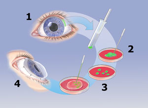
2. In vitro isolation and proliferation of limbal tissue on top of human amniotic membrane for 3-4 weeks.
3. Growth of limbal tissue of 2-3 cm2 in diameter.
4. Removal of the damaged epithelial tissue before implantation of the cultured epithelial limbal tissue.
Basic principles ||
In the cornea usually exists a natural exchange of epithelial cells, where the most superficial elements are detached from the epithelium and are replaced by basal cells.
For the perpetuation of the system there must be necessarily epithelial-corneal "stem" cells, which reside mainly in the Vogt Palisade in the epithelial-limbal region.
The re-epithelisation of the corneal surface by conjunctival keratinized cells, invariably produces chronic inflamation, permanent epithelial defects, formation of scar and neovascularization that causes defects of the vision.
The deficiency of corneal epithelial "stem" cells might originate in the destructive loss of limbal "stem" cells like in the dysfunction of limbal stroma or in inherited pathologies like epidermal dysplasia or acquired by multiple causes.
Within the most frequent acquired pathologies are lesions produced by thermal and
chemical burns, severe microbial infections which involve the limbus, lesions by radiation, multiple surgeries or cryotherapy, drug toxicity (mitomycin, 5-flurouracil), Stevens Johnson syndrome and keratopathies induced by contact lenses.
Different limbal stromal pathologies can also give rise to the deficiency of "stem" cells.
These lesions again can have genetic origins like aniridia and keratitis associated to multiple deficiencies of the endocrine system.
The ulcerated peripheral keratitis (Mooren´s ulcer), neurotrophic keratopathy, pterygium, chronic limbitis, drug toxicity and radiation, are some of the acquired causes which can produce stromal lesions.
Recently, various surgical procedures have been reported with the intention to repopulate the limbal surface with functional "stem" cells. This can be achieved by the transplantation of limbal epithelial tissue from the healthy contralateral eye of the same patient (autologous transplantation) or donor material (allogenic transplantation).
At present, the ex vivo expansion of limbal epithelial "stem" cells is performed on top of human amniotic membrane. This membrane has been used for many years in a wide range of ocular surgeries and proved to be very useful for the treatment of severe pterygium, chemical burns, Stevens Johnson syndrome and other diseases. This characteristic makes this material a very adequate substrate to achieve proliferation of epithelial limbal cells.
In Italy, there are registered more than 1,000 procedures of this type, as well as in USA and Europe, representing a valid alternative for the repair of damaged cornea.
|| printable version

Agrelo
3038 - C.P. 1221/Buenos Aires / Argentina - Teléfono/Fax: (+54-11)
4932-7068/4956-1579 - mail: info@cellprep.com
(c) 2000 CellPrep S. A. Todos los derechos reservados.
(c) 2000 CellPrep S. A. Todos los derechos reservados.