


>> culture of chondrocytes for cartilage repair
|
Location: preferably
from the medial trochlea, it could also be obtained from the lateral
or the intercondilean gap Size: at least 2 cylinders of 4 mm diameter, weight 200 mg Transport: sterile medium provided by CellPrep Second surgical procedure by arthrotomy (Illustration of a case) |
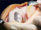
| 1.
Grade IV defect of articular cartilage of the left medial femoral
condyle |
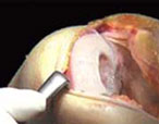 |
| 2. Dissection of the
defect: it should be sectioned carefully with an oval or circular
configuration, to avoid penetration of the subchondral bone |
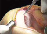 |
| 3. Debridement: the
non-adherent and degenerated cartilage should be removed completely.
The walls of the defect should be perpendicular to the subchondral
bone. |
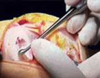 |
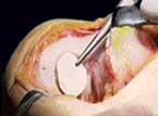
| 4. Creation of a template:
measurement of the defect size by means of a template of metal foil. |
 |
| 5. Dissection of the
periosteum: the periosteum which is extracted from the front surface
of the tibia during the procedure is excised according to the size
of the template. |
 |
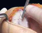
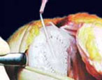
| 6. Procedure of suture:
the periosteum is sutured to the healthy neighbouring cartilage
tissue by using the inside to outside technique. |
 |
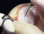
| 7. The sites of suture
and the borders of the union between the periosteum and the neighbouring
cartilage are sealed using fibrin glue. |
 |
| 8. Test of the integrity
of the cartilage-periosteum union by injection of saline solution
or sterile Ringer. As soon as the integrity of the union is proved and the test-serum is removed, the suspension of the cultured chondrocytes is injected. |
 |
|| printable version

Agrelo
3038 - C.P. 1221/Buenos Aires / Argentina - Teléfono/Fax:
(+54-11) 4932-7068/4956-1579 - mail: info@cellprep.com (c) 2000 CellPrep S. A. Todos los derechos reservados. |
||||